Case 1
Patient information: a 57-year-old Tunisian patient (north Africa) presented to the neurology department with history of difficulty in walking with weakness and stiffness in both lower limbs. The weakness gradually progressed over the past 6 months. There was no history of sensory impairment and symptoms of bowel and bladder involvement were absent.
His medical history revealed liver cirrhosis related to Budd–Chiari syndrome with partial thrombosis of the inferior vena cava and superior right hepatic vein. Etiological investigation was negative and the patient was put on anti-vitamin K.
Clinical finding: at admission he did not complain of headaches, fever, or fasciculations. Systematic examination was unremarkable and neurological examination found a spastic paraparesis with scissor walk. In addition, there is quadripyramidal syndrome, especially in lower limbs with brisk deep tendon reflexes, and bilateral Babinski sign. There was no disturbance of superficial or deep sensitivity and cranial nerves examination was normal.
Diagnostic assessment: laboratory investigations revealed bicytopenia, hypocholesterolemia, liver transaminases were normal (Alanin aminotransferase (ALT), Aspartate aminotransferase (AST) (15/12 IU/l), total bilirubin/direct (10/5). Cerebrospinal fluid (CSF) examination without abnormalities (Table 1). Patient’s serum was no reactive to hepatitis A, B, C, lyme, and human immunodeficiency virus (HIV). Tests for a autoimmune diseases were negative, especially for antinuclear (AAN), Anti Neutrophil Cytoplasmic Antigen (ANCA), and aquaporin 4 antibodies.
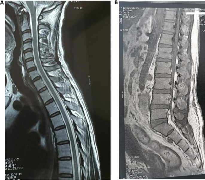
Medullar magnetic resonance imaging (MRI) noted the presence of multiple disco-vertebral degenerative phenomena staged on a narrow bipolar spinal canal without spinal cord compression or root conflict (Fig. 1).
Cerebral MRI T2 showed a bilateral and symmetric hypersignal bipallidal (Fig. 2).
Diagnosis: hepatic myelopathy caused by a spontaneous portocaval shunt in the context of Budd–Chiari syndrome.
Therapeutic interventions: the patient received conservative therapies with antispastic drugs and motor reeducation.
Patient perspectives: a stabilization of the handicap.
Informed consent: applicable.
Case 2
Patient information: a 37-year-old Tunisian patient (north Africa) presented to the neurology department for progressive walking disorders and sensation of heavy legs without sensitivity disorders. Her medical past history included Hodgkin’s lymphoma treated with radiochemotherapy in 2013 and viral B cirrhosis since 2017.
Clinical finding: neurological examination showed spastic paraplegia. Quadripyramidal syndrome especially in lower limbs with lively diffuse tendon reflexes, a bilateral Babinski sign, and clonus of the lower limbs. No sensitivity disorders or vesicosphincter disorders were noted.
Diagnostic assessment: her biological assessment showed hepatic failure with thrombocytopenia: 91,300, low TP: 35.5%; total bilirubin: 50 IU/l; low albumin level at 21.8 g/dl; Alanin aminotransferase (ALT), Aspartate aminotransferase (AST): 61/46 IU/l and hyperammoniemia (81.11 mmol/l) (Table 1).
Medullary MRI was normal and cerebral MRI showed T2 hypersignals of the pallidums.
Diagnosis: as infectious, autoimmune, paraneoplasic, and compressive cause of myelopathies were eliminated, the diagnosis of hepatic myelopathy was made
Therapeutic interventions: the patient was treated with baclofen, vitamins, and nutritional supplementation, as well as motor rehabilitation and listed for liver transplantation.
Patient perspectives: on follow-up after 3 months, we noted a partial improvement.
Patient consent: applicable.


Add Comment