A 42-year-old South Asian male, a father of two, presented to the emergency treatment unit of a District General Hospital in Sri Lanka following deliberate self-ingestion of S following an attempted suicide. No other toxins were found in the vicinity. His past medical, surgical, and allergy history had been insignificant. Admission was within 90 min following the ingestion. On admission, his airway was patent with equal bilateral air entry without added sounds, respiratory rate was 16 breaths per minute with a shallow breathing pattern, and capillary O2 saturation was 92% in room air. The pulse rate was 110 bpm; capillary refill was less than 2 s with a blood pressure of 140/85 mmHg. GCS was 11/15 (E-3, V-3, M-5), and bilateral pupils were 3 mm in size and equally reacted to light. Fundoscopy did not reveal papilledema. The capillary blood glucose level was 130 mg/dl.
The patient was managed symptomatically with face mask O2 of 10 l/min. He was catheterized, and an input–output chart was generated. A 16G NGT was inserted, and he was treated with activated charcoal. Over 90 min, 3% saline 150 ml was given to alleviate possible cerebral edema. Blood was sent for investigations including venous blood gas. The latter yielded normal acid–base status, electrolytes, and lactate. An electrocardiogram revealed sinus tachycardia with a rate of about 120 bpm. X-ray of the chest was normal. The patient’s GCS gradually deteriorated, and simultaneously airway patency was reduced with stridor. He was intubated under direct laryngoscopy following rapid sequence induction with IV propofol 2 mg/kg and IV suxamethonium 2 m/kg and cricoid pressure by an anesthetist. The pharynx appeared inflamed. Intravenous dexamethasone 8 mg stat dose was administered. Arterial blood gas after the intubation revealed a pH of 7.43, PCO2 27 mmHg, PO2 241 mmHg with lactate of 4.3 mmol/l and base excess of −5 mmol/l. A 500 ml 0.9% saline bolus was administered, and the maintenance fluid was increased to 120 ml/h.
The patient was admitted to the intensive care unit (ICU) for further management and monitoring. His initial blood investigations are illustrated in Table 1.
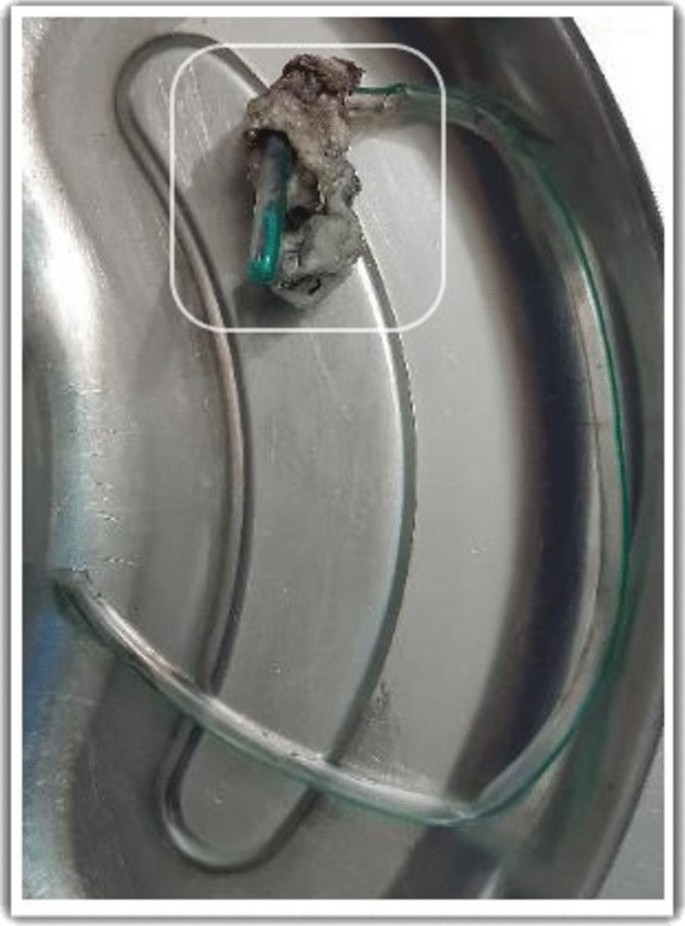
At the ICU, the patient was sedated and paralyzed to carry out lung and neuroprotective ventilation. Supportive care was given with stress ulcer and thromboprophylaxis. ENT referral was performed to assess the laryngeal and pharyngeal areas under fiber-optic laryngoscopy (FOL), and the findings were compatible with chemical laryngitis with mildly inflamed glottis, epiglottis, mildly edematous subglottic area, and vestibule of the larynx. Regular intravenous dexamethasone was administered. The patient was kept nil by mouth on the first 2 days, and NG feeding commenced after 48 h. His liver enzymes gradually dropped to normal levels. On day 3, sedation was stopped. The GCS had improved to 10/10. Successful extubation of the trachea was performed. Repeat FOL revealed that the laryngopharyngeal edema had now resolved. The patient appeared anxious and did not tolerate the NGT. As the patient was tolerating clear fluids via the oral route, the decision was made for its removal. Removal failed as the NGT was found to be lodged in the nasopharynx and attempts at pushing it back toward the oropharynx were unsuccessful. The patient became extremely anxious not responding to routine doses of sedatives. For the safety of the patient and the staff, he was reintubated and sedated. Subsequently, the NGT was removed by the ENT surgeon; the procedure was mildly traumatic with mild bleeding from the nasopharynx. The nasopharynx was packed with an adrenaline-soaked gauze. A Foley catheter was inserted, and the bulb was kept inflated for tamponade effect. The distal end of the NGT had formed a clump after reacting with S, which had led to the initial failure in removal (Fig. 1). The ENT team’s advice was to keep the pack for 48 h, after which the ENT team could remove the pack and the catheter. FOL was repeated, and inflammation of the laryngopharyngeal area had improved with the absence of significant bleeding. The leak test was repeated and found to be positive.
The nasal pack was removed after 48 h, and the patient’s trachea was extubated. Oral feeding was gradually commenced. He was discharged to the ward on day 5. A psychiatry referral was done at the ward, and upper gastrointestinal (GI) endoscopy was arranged at a tertiary care center in 2 weeks. Toxicology studies were not carried out as these were not available freely in this low-resource setting.


Add Comment