Case 1
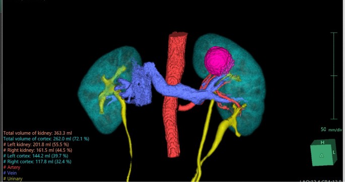
A 45-year-old Vietnamese male patient was incidentally discovered to have a left kidney mass during a routine health check. The patient had a history of hypertension and no other complaints. The CT scan revealed a distinct, confined mass lesion at the upper pole of the left kidney, measuring 32 × 33 × 35 mm, suggestive of RCC. The left kidney was entirely duplex on imaging. We utilized the Fujifilm’s Synapse® Artificial Intelligence (AI) Platform to reconstruct a 3D image of both kidneys and clearly observed the fully duplicated left kidney along with the tumor at the upper pole. Using Tc-99 m DTPA isotope scan, the estimated split renal function of the left kidney was 52.6%, with a glomerular filtration rate of 40.3 ml/minute.
The diagnosis in this case is cT1aN0M0 hilar renal tumor on the entirely duplicated left kidney, Renal score was 9ah. The patient underwent a partial nephrectomy of the left kidney with the assistance of the da Vinci robotic system. During the surgery, we exposed and observed the anatomical structure of the left duplex kidney as seen in the 3D reconstructed image (Fig. 1). The renal tumor appeared distinctly with well-defined borders and was located at the upper pole of the left kidney. We clamped the left renal artery using a Bulldog clamp and proceeded to perform an enucleation of the renal tumor. The warm ischemia time was 23 minute, estimated blood loss was approximately 50 ml, and the total surgical time was 140 minute.
The patient was discharged after 3 days with no postoperative complications observed. The histopathological results confirmed renal cell carcinoma (RCC), with no tumor cells found at the surgical margins, indicating clear resection (Fig. 2). One-month follow-up, the renal function was preserved without change in the serum creatinine level.
Case 2
A 54-year-old Vietnamese female patient was incidentally found to have a right kidney mass during a routine health examination. The CT scan imaging was strongly suggestive for a solid renal mass, likely renal cell carcinoma (RCC). Preoperative tests showed normal results, and both kidneys demonstrated comparable function. The left kidney had a glomerular filtration rate of 50.9 ml/minute, constituting 50.1% as determined by the Tc-99 m DTPA renal isotope study.
With the assistance of Fujifilm’s Synapse® AI Platform, we obtained detailed images of the tumor anatomy, including the blood vessels supplying the tumor. Additionally, the AI platform helped identify the presence of bilateral duplicated kidneys, a detail that was initially overlooked in the conventional CT scan (Fig. 2).
The patient was diagnosed with RCC, clinically staged as cT1bN0M0, and had a RENAL score of 9x. A partial nephrectomy of the right kidney was performed using the da Vinci robotic system. During the surgery, distinct images of the two collecting systems were observed, clearly depicted in the 3D reconstructed image using AI (Figs. 3 and 4). The tumor was prominently located at the lower pole, showing well-defined borders with the normal parenchyma and no invasion into the underlying collecting systems. Examination of the renal hilum revealed a perfect match between the actual branching pattern of the blood vessels and the image generated by Fujifilm’s Synapse® AI Platform.
We clamped the renal artery using a laparoscopic Bull-dog clamp and proceeded to perform an enucleation of the renal tumor. The warm ischemia time was 28 minute, estimated blood loss was approximately 60 ml, and the total surgical time was 150 minute.
The patient was discharged after 4 days with no postoperative complications observed. The histopathological results confirmed renal cell carcinoma (RCC), with no tumor cells found at the surgical margins (Fig. 5). Furthermore, the serum creatinine level was unchanged one month postoperatively.


Add Comment